EN

CIENCIA


viernes, 27 de agosto de 2010

TAURINE

Richard Marsh and Paul May
Bristol University, UK

Gives you Wings?

Perhaps the most famous use of taurine in recent years has been in the energy drink Red Bull. Originating on college campuses and in nightclubs in Austria, the product quickly spread. The drink's marketing leans heavily on its unusual ingredient, and so integral is the substance to the brand that even the name derives from it - taurus is Latin for bull (as taurine was first isolated from ox bile in 1827). Red Bull promises that the taurine, along with caffeine and, in its original form, a great deal of sugar, will help to 'vitalise body and mind'.

Red Bull LogoBut what effect does the taurine in the drink actually have on the body? Research into this area is limited, but some researchers are sceptical [1]. They postulate that the effects of the drink can be attributed almost entirely to its caffeine content, and suggests that the increased effect compared to a cup of coffee (which contains a similar amount of caffeine) is only due to the temperature of the two drinks. A cold cup of coffee should have essentially the same physiological effect. Also they suggest a psychosomatic element to the drink's effects. In terms of the muscular effect of taurine, while it is possible that ingesting extra taurine would increase the force generation of the muscles no studies have been completed to demonstrate this and, given the naturally occurring levels of taurine in the body, it seems unlikely the dose added by Red Bull would cause a significant enough increase to have noticeable effects on the muscles.

However, other researchers' findings support the company's claims. Seidl et al [2] report a double blind placebo controlled survey on a number of university students who demonstrated increased motor response and alertness compared to those receiving the placebo. Unfortunately the test did little to determine which of the drink's ingredients were responsible for the effects.

So what is taurine?

Although by the strictest definition, taurine is not an amino acid as it does not contain a carboxylic acid COOH group [3], it is generally referred to as one in published literature [4, 32,33,37]. At pH 7, the molecule exists as a zwitterion, in which the NH2 end of the molecule exists as NH3+ and the opposite end as SO3-. This gives the molecule two polar ends and a non-polar carbon chain centre, allowing for a great many possible binding interactions. Unusually for a biological compound, taurine does not exhibit chirality.

Taurine structure - click for 3D VRML structureTaurine - space fill model
Taurine - (2-aminoethane sulphonic acid) [1]

Its Role in Biology and Pharmacy

Taurine is synthesised by the human body, primarily in the liver by oxidation followed by decarboxylation of the amino acid cysteine.

Biosynthesis of taurine
The biosynthesis of taurine from cysteine in the liver [5].

Taurine is one of a group of organic compounds which has been formed in experiments designed to simulate early-Earth conditions along with electrical discharges to simulate lightning. This result is often cited as evidence for the ease with which sulfur-containing biological molecules could be naturally synthesised under prebiotic conditions [6]. The conclusion is somewhat controversial, however, as the reaction occurred because of the presence of large concentrations of methane and ammonium hydroxide, the abundance of which on prebiotic Earth is questionable [7].

With a few exceptions, liver-synthesised taurine is not incorporated into polypeptides but is found free, lending it a unique set of properties, including a separate intercell transport mechanism and an independence from protein synthesis and catabolism [8]. Taurine has been linked to a wide range of bodily functions and Stapleton et al (1998) list roles in "osmoregulation, antioxidation, detoxification and stimulation of glycolysis and glycogenesis". The synthesis pathway of taurine is especially active in the early stages of life, and taurine is found in breast milk, suggesting that it is particularly vital at this stage. Taurine also plays a role in muscle contraction, where it enhances the ability of the muscles to generate force by catalysing the uptake and release of calcium ions [9].

Experiments on rats have also yielded evidence of a very substantial role in detoxification in the liver - Waters et al [10] report that high doses of taurine administered before or soon after ingestion of an overdose of paracetemol protected the livers of rats from hepatotoxcity and serious liver damage from the drugs. Although the idea has yet to progress to any form of human trial, the prophylactic and therapeutic benefits of the substance in cases of paracetemol overdose are promising .

A blind cat - a result of taurine deficiency?In fact, several areas of medical research are interested in taurine's pharmaceutical potential. For example, phase two clinical trials are currently underway at McLean Hospital in Massachusetts using taurine as an anti-manic agent to stabilise the mood of patients with bipolar disorder [11]. Tsuboyama-Kasaoka et al [12] also postulate a link between taurine deficiency and obesity in humans, showing that in mice, raising taurine levels in the body led to a greater resting rate of energy usage and less build-up of adipose tissue. The paper therefore suggests that taurine supplements may help prevent obesity.

Taurine in animals

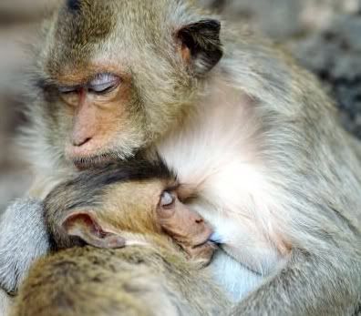
Taurine is necessary for normal skeletal muscle functioning in humans and mice [13]. Cats cannot synthesise taurine, and so need it added as supplements in their diets. Without it, they suffer conditions such as blindness, hair loss and tooth decay. Taurine is also essential in the early development of many types of perching birds. Parent birds with new offspring often seek out spiders (which are rich in taurines) with which to feed their young. so, in this case, taurine really does give them wings!

References

1. Kim W., "Debunking the Effects of Taurine in Red Bull Energy Drink", Nutrition Bytes, 9, (2003) 6.

2. Seidl R. et al, "A Taurine and Caffeine-containing drink stimulates cognitive performance and well-being", Amino Acids, 19, (2000) 635.

3. Sharp D., Dictionary of Chemistry (Abridged), 3rd Edition, Penguin Books, 2003.

4. Stapleton P. et. al, "Host defense - a role for the amino acid taurine?", J. Parenteral and Enteral Nutrition, 22, (1998) 42.

5. Moss G., "Taurine Biosynthesis", http://www.chem.qmul.ac.uk/iubmb/enzyme/reaction/misc/taurine.html, Queen Mary University of London, 1992.

6. Ferris J., "Chemical markers of Prebiotic Chemistry in Hydrothermal systems", in Origin of Life and Evolution of Biospheres V.22 Numbers 1-4, ed. N Holm, Springer, Netherlands, Jan. 1992, p.120.

7. Wigley T., Nature, 291, (1981) 213.

8. Chiarla C. et al, "The Relationship between Plasma Taurine and Other Amino Acid Levels in Human Sepsis", J. of Nutrition, 130, (2000) 2222.

9. K. Harrison, "Taurine", http://www.3dchem.com/molecules.asp?ID=22, (2002).

10. Waters E. et al, "Role of taurine in preventing acetaminophen-induced hepatic injury in the rat", Am. J. Physiol. Gastrointest. Liver Physiol., 280, (2001) 1274.

11. Murphy B. et al, "Taurine as an Anti-Manic Agent", (2005) http://www.clinicaltrials.gov/ct/show/NCT00217165

12. Tsuboyama-Kasaoka et al, "Taurine (2-Aminoethanesulfonic Acid) Deficiency Creates a Vicious Circle Promoting Obesity", Endocrinology, 147, (2006) 3276.

13. Wikipedia - taurine

lunes, 9 de agosto de 2010

In a Video Game, Tackling the Complexities of Protein Folding

In a match that pitted video game players against the best known computer program designed for the task, the gamers outperformed the software in figuring out how 10 proteins fold into their three-dimensional configurations.

Proteins are essentially biological nano-machines that carry out myriad functions in the body, and biologists have long sought to understand how the long chains of amino acids that make up each protein fold into their specific configurations.

In May 2008, researchers at the University of Washington made a protein-folding video game called Foldit freely available via the Internet. The game, which was competitive and offered the puzzle-solving qualities of a game like Rubik’s Cube, quickly attracted a dedicated following of thousands of players.

The success of the Foldit players, the researchers report in this week’s issue of Nature, shows that nonscientists can collaborate to develop new strategies and algorithms that are distinct from traditional software solutions to the challenge of protein folding.

The researchers took pains to credit the volunteers who competed at Foldit in the last two years, listing “Foldit players” at the end of the report’s author list and noting that more than 57,000 players “contributed extensively through their feedback and gameplay.”

Zoran Popovic, a computer scientist at the University of Washington who was a lead author of the paper, said, “If things go according to plan, not too long from now, such massive author lists should be commonplace.” Foldit begins with a series of tutorials in which the player controls proteinlike structures on a computer display. In the game, as structures are modified, a score is calculated based on how well the protein is folded. Players are given a set of controls that let them do things like “shake,” “wiggle” and “rebuild” to reshape the backbone and the amino acid side shapes of a specific protein into a more efficient structure.

A list of top scores for each puzzle is posted so that players can compare their results. Players may also collaborate in teams, tracking progress on a separate list of group scores.

The protein-folding problem can be solved by computers using statistical and related software algorithms, but it takes an immense amount of processing power.

“The problem is that these proteins are far, far more complex than a robotic arm, and can ‘fold’ in time frames measured in billionths of a second,” Duke Ferris, founder of the GameRevolution Web site, wrote recently. “It’s like trying to solve a million-sided Rubik’s Cube while it also spins at 10,000 r.p.m. And that’s for just one ‘fold.’ ”

In a comparison involving 10 separate protein-folding puzzles, video game players matched the results generated by software solutions in three of the puzzles, outperformed them in five cases and found significantly better solutions in two others, according to the scientists.

In addition to the acuity of human pattern-recognition skills, the researchers noted that players outperformed the best software tools in other ways as well, writing: “Humans use a much more varied range of exploration methods than computers. Different players use different move sequences, both according to the puzzle type and throughout the duration of a puzzle.”

The Foldit project was inspired by the volunteers who were contributing the downtime on their home computers to power a protein-folding program called Rosetta@home. The computer donors could see the progress of the program on their screens, and they began to note inefficiencies in the software’s folding approach. That led the scientists to look for ways to systematically harness the skills of the human volunteers.

Two New Paths to the Dream: Regeneration

By NICHOLAS WADE Published: August 5, 2010

Two research reports published Friday offer novel approaches to the age-old dream of regenerating the body from its own cells.

Animals like newts and zebra fish can regenerate limbs, fins, even part of the heart. If only people could do the same, amputees might grow new limbs and stricken hearts be coaxed to repair themselves.

But humans have very little regenerative capacity, probably because of an evolutionary trade-off: suppressing cell growth reduced the risk of cancer, enabling humans to live longer. A person can renew his liver to some extent, and regrow a fingertip while very young, but not much more.

In the first of the two new approaches, a research group at Stanford University led by Helen M. Blau, Jason H. Pomerantz and Kostandin V. Pajcini has taken a possible first step toward unlocking the human ability to regenerate. By inactivating two genes that work to suppress tumors, they got mouse muscle cells to revert to a younger state, start dividing and help repair tissue.

What is true of mice is often true of humans, and although scientists are a long way from being able to cause limbs to regenerate, the research is attracting attention. Jeremy Brockes, a leading expert on regeneration at University College London, said the report was “an excellent paper.” Though there is a lot still to learn about the process, “it is hard to imagine that it will not be informative for regenerative medicine in the future,” he said.

In recent years, most research in the field of regenerative medicine has focused on the hope that stem cells, immature cells that give rise to any specific type of cell needed in the body, can somehow be trained to behave as normal adult cells do. Nature’s method of regeneration is quite different in that it starts with the adult cells at the site of a wound and converts the cells to a stemlike state in which they can grow and divide.

The Stanford team has taken a step toward mimicking the natural process. “What I like is that it’s built on what’s happening in nature,” Dr. Blau said. “We mammals lost this regenerative capacity in order to have better tumor suppression, but if we reawaken it in a careful way we could make use of it in a clinical setting.”

Dr. Pomerantz, a clinician, hopes the technique can be applied to people, though many more animal experiments need to be done first. “We have shown we can recapitulate in mammalian cells behavior of lower vertebrate cells that is required for regeneration,” he said. “We would propose using it in amputations of a limb or part of a limb or in cardiac muscle.” After a heart attack, the muscle cells do not regenerate, so any method of making them do so would be a possible treatment.

Interfering with tumor suppressor genes is a dangerous game, but Dr. Pomerantz said the genes could be inhibited for just a short period by applying the right dose of drug. When the drug has dissipated, the antitumor function of the gene would be restored.

Finding the right combination of genes to suppress was a critical step in the new research. One of the two tumor suppressor genes is an ancient gene, known as Rb, which is naturally inactivated in newts and fish when they start regenerating tissue. Mammals possess both the Rb gene and a backup, called the Arf gene, which will close down a cancer-prone cell if Rb fails to do so.

The Stanford team found that newts did not have the Arf backup gene, which mammals must have acquired after their lineage diverged from that of amphibians. This suggests that the backup system “evolved at the expense of regeneration,” the Stanford researchers say in Friday’s issue of Cell Stem Cell.

The Stanford team shut off both Rb and Arf with a chemical called silencing-RNA and found the mouse muscle cells started dividing. When injected into a mouse’s leg, the cells fused into the existing muscle fibers, just as they are meant to.

The Stanford researchers have learned how to block two genes thought to inhibit the natural regenerative capacity of cells, but it is somewhat surprising that the regenerative mechanism should still exist at all if mammals have been unable to use it for 200 million years. “One school of thought is that regeneration is a default mechanism and doesn’t require its own program,” Dr. Pomerantz said.

Dr. Brockes believes that this is true in part. Regeneration “depends on a largely conserved cellular machinery,” he said, meaning that it is present in all animals. The machinery comes into play in wound healing and tissue maintenance. But specific instances of regeneration, like regrowing a whole limb, are invoked by genes specific to various species. He has found a protein specific to salamanders that coordinates regrowth of a salamander limb.

If the regeneration of a whole limb is a special ability that salamanders have evolved, then humans would not have any inherent ability to do the same. “I would beware of suggesting that this sort of manipulation is capable of unlocking ‘the newt within,’ ” Dr. Brockes said.

A second, quite different approach to regenerating a tissue is reported in Friday’s issue of Cell by Deepak Srivastava and colleagues at the University of California, San Francisco. Working also in the mouse, they have developed a way of reprogramming the ordinary tissue cells of the heart into heart muscle cells, the type that is irretrievably lost in a heart attack.

The Japanese scientist Shinya Yamanaka showed three years ago that skin cells could be converted to embryonic stem cells simply by adding four proteins known to regulate genes. Inspired by Dr. Yamanaka’s method, Dr. Srivastava and his colleagues selected 14 such proteins and eventually found that with only three of them they could convert heart fibroblast cells into heart muscle cells.

To make clinical use of the discovery, Dr. Srivastava said he would need first to duplicate the process with human cells, and then develop three drugs that could substitute for the three proteins used in the conversion process. The drugs could be loaded into a stent, a small tube used in coronary bypass operations. With the stent inserted into a heart artery, the drugs would convert some of the heart’s tissue cells into heart muscle cells.

Some researchers hope that with Dr. Yamanaka’s method of turning skin cells into embryonic stem cells, those stem cells can be converted into usable heart muscle cells. One problem with this approach is that any unconverted embryonic stem cells may form tumors. Dr. Srivastava’s method sidesteps this problem by avoiding the stem cell stage.

martes, 3 de agosto de 2010

2 August 2010
BIOCHEMIST EVOLUTION
microRNA protects red blood cells from free radicals

Paediatric researchers have discovered a new biological pathway in which small segments of RNA, called microRNA, help protect red blood cells from injury caused by free radicals.

The microRNA seems to have only a modest role when red blood cells experience normal conditions, but steps into action when the cells are threatened by oxidant stress.

Led by haematologist Mitchell Weiss of The Children’s Hospital of Philadelphia, the current study describes how a particular microRNA fine-tunes gene activity by acting on an unexpected signalling pathway.

The study appears in the August 1 issue of the journal Genes & Development, simultaneously with a similar study of microRNAs and red blood cells by a University of Texas team led by Eric Olson. The two studies reinforce each other, said Weiss.

MicroRNAs are single-stranded molecules of ribonucleic acid (RNA) averaging only 22 nucleotides long. Scientists estimate that 500 to 1000 microRNAs exist in the human genome. First characterized in the early 1990s, they received their current name in 2001. Over the past decade, scientists have increasingly recognized that microRNAs play a crucial role in regulating genes, most typically by attaching to a piece of messenger RNA and blocking it from being translated into a protein, but many details remain to be discovered.

“Although microRNAs affect the formation and function of most or all tissues, for most microRNAs, we don’t know their precise mechanisms of action,” said Weiss. “In this case we already knew this microRNA, called miR-451, regulates red blood cells in zebrafish and mice, and because it is highly conserved in evolution, we presume it operates in humans as well. But its functional roles were poorly understood.”

By investigating how microRNAs influence red blood cell development, Weiss and colleagues aimed to understand how such development goes wrong in haemolytic anaemia, in which red blood cells are destroyed in large numbers, or in disorders of abnormal blood cell production. The current study used knockout mice — bioengineered animals in which the miR-451 gene was removed and could not function.

They found that preventing the activity of miR-451 produced only modest effects — mild anaemia in the mice — but when the team subjected mice to oxidant stress by dosing them with a drug that produces free radicals, the mice had profound anaemia. The oxygen radicals attacked haemoglobin, the iron-carrying molecule in red blood cells.

“This is a common theme in microRNAs — frequently, they don’t play a central role during tissue formation or normal conditions, but they have a strong protective effect when an organism is stressed,” said Weiss. “Over evolutionary time, red blood cells have evolved ways to protect themselves; one of those ways is the action of microRNA.”

Weiss’s team found that miR-451, acting through intermediate steps on a signalling pathway, affects a key protein, FoxO3. As a transcription factor, FoxO3 regulates hundreds of genes; in this case, FoxO3 stimulates specific genes that protect red blood cells from oxidant stress. The knockout mice in this study, having lost miR-451’s function, showed impaired FoxO3 activity, and less ability to protect their red blood cells.

The regulatory pathway seen here, Weiss added, may have medical implications beyond blood cell development. “This finding does not have immediate clinical application for patients with blood diseases, but it sheds light on how microRNAs fine tune physiological functions in different contexts,” said Weiss. FoxO3 regulates anti-oxidant functions in heart cells and also acts as a tumour suppressor, so miR-451 may have an important role in heart protection and in fighting cancers. “Further studies may broaden our knowledge of how this microRNA may defend the body against disease,” he added.

viernes, 25 de junio de 2010

DIABETES AND PREGNANCY 2011


Diabetes & Pregnancy Meeting Salzburg, Austria March 24-26, 2011

By embryology


The 6th International Symposium on Diabetes & Pregnancy (DIP) Salzburg, Austria March 24-26, 2011

The field of diabetes and pregnancy has come of age. From the conception of the terminology ‘gestational diabetes’ and ‘diabetes in pregnancy’ to the creation of an entire subspecialty, this Symposium documents the ‘gestation’ of the field.
This Symposium not only documents the past 80+ years of progress in the field of diabetes and pregnancy, but also presents the most up-to-date tools, techniques and management protocols to ensure the optimal outcome of pregnancies complicated by diabetes. In addition, the areas that remain controversial, such as screening and diagnosis, will be discussed in detail to enable participants to come to an opinion while waiting for the evidence to validate many of the expert opinions presented.
The latest theories and literature on the immunology of Type 1 diabetes will also be included, giving us hope that the near future holds the answers to prevention of this disease. Until there is a cure for diabetes, we must continually take on the weight of astutely diagnosing diabetes and treating all pregnant women who are at risk of an untoward outcome of pregnancy.
The faculty is comprised of renowned professionals and practitioners, who will present based on evidence as well as clinical experience.
In addition, this Symposium provides an international approach to enhancing the quality of care for women with diabetes in pregnancy, and answers for the clinician to enable delivery of optimal care for all pregnancies complicated by diabetes.

miércoles, 9 de junio de 2010

2010 International Year of Biodiversity

The UN has designated 2010 the 'International Year of Biodiversity’. To mark and celebrate this, the Society of Biology is organising a photographic competition. Entrants may submit one image per category. Entrants are free to interpret how they capture photographically images that celebrate, explore, comment on or reveal aspects of biodiversity from around the world.

Categories
  • Land – biodiversity associated with land
  • Air – biodiversity associated with the air
  • Water – biodiversity associated with water
  • Close up – biodiversity unseen by the naked eye
Prizes
  • Best in Show – £1,000
  • 1st prizes in each category – £250 in cash and Park Cameras vouchers
  • Top Young Photographer winner – £250 worth of vouchers
  • There will be additional prizes for runners up and certificates for ‘highly commended’ entries
The competition is free, open to all and will run between 20 April - 20 August 2010. Please review the competition rules before entering, details of the rules and entry forms are located to the right. Please contact photocomp@societyofbiology.org with any questions

Annual Symposium - Recent advances in membrane biochemistry

5—7 January 2011

Robinson College, Cambridge, UK


Earlybird registration deadline: 3 December 2010.

Registration will be available shortly.


Abstract submission deadline: 26 October 2010.

Abstract submission will be available shortly.


There are Student Travel Grants available for this meeting.

Oral communication slots are available at this meeting. All attendees, particularly young researchers, are invited to submit a poster abstract for consideration as an oral communication.


The proceedings of the meeting will be published as a stand-alone issue of Biochemical Society Transactions (online and in print) and a stand-alone volume of the online Symposia Series.

Symposium background
This symposium will highlight some of the recent advances in molecular membrane biochemistry. Membrane biology underpins a vast array of life processes such as bioenergetics, signalling and transport. In the last few years advances in this field has led to the development of large scale expression and purification of mammalian transmembrane proteins. This has then been paramount to enabling efficient crystallization and elucidation of the 3-dimensional structures of transmembrane proteins to occur.

In addition, other biophysical techniques have also been employed to further understand membrane protein structure, conformations, and interactions with other membrane components.


This symposia will focus on a number of key areas of current research advances, which will form the basis of 5 sessions:

1) Membrane protein expression and 3-dimensional structural analysis.


2) Examples of expression / structure analysis of membrane proteins which are of current interest.


3) Computer modelling of membrane protein structure, membrane protein folding and dynamics.


4) Lipid-protein interactions.

5) Membrane proteins in disease.

miércoles, 26 de mayo de 2010

Big Bang in the protein universe

Researchers of the Centre for Genomic Regulation (CRG) demonstrate evidence in support of the common ancestry of life thanks to a new computational approach to study protein evolution.

The study, published in Nature, reveals that protein evolution has not reached its limit and it is still continuing. At the same time, it provides us new information on why this evolution is so slow and conservative showing that protein structures are more evolutionary plastic than previously thought.

Almost 100 years ago Edwin Hubble observed that distant galaxies are moving away from Earth faster than those that are closer. This relationship between distance and velocity is widely cited as evidence of the origin of the Universe from a Big Bang. Researchers at the Centre for Genomic Regulation copied his approach to investigate the divergence between protein sequences.

“We wanted to know if the divergent evolution between proteins was still proceeding. Today, we can find proteins that are still similar after almost 3.5 billion years of evolution. Our study showed that their divergence continues with these proteins becoming more and more different despite their incredible level of conservation”, said Fyodor Kondrashov, principal investigator of the project and leader of the Evolutionary Genomics group at the CRG.

The work done by Fyodor Kondrashov and Inna Povolotskaya goes beyond similarity studies and discusses the evolution of proteins from the view of evolutionary dynamics, offering a new perspective on how protein structures are maintained in evolution. “In the same way that Hubble’s observations led to an understanding of the past and the future of our universe, using his approach at a molecular level we get a similar overview that gives us the ability to analyze evolutionary dynamics and get a broad prediction of the possible changes to the proteins in the future”, says Inna Povolotskaya, first author of the work and responsible for obtaining and analyzing all data.

Proteins are formed through combinations of amino acids, with only 20 types of amino acids are available to form a particular protein. To obtain the data for their study, the CRG researchers have compared proteins sequences from different species that were available in GenBank, a public database of genetic information. Comparing these sequences the authors measured the distance of proteins from each other and devised a method for measuring how fast the proteins are accumulating different changes. Thus, they could replicate Hubble’s approach by correlating the distance between the proteins with the rate of their divergence. The result indicates that even the most distantly-related proteins are still accumulating differences.

The study shows how new techniques of bioinformatics and computational analysis can also expand knowledge at a molecular level. “Our work is a good example of how we can learn new and very fundamental things just by analyzing a larger volume of data that can be obtained by one experimental laboratory”, says Kondrashov.

Most changes in a protein are deleterious because they somehow disrupt its structure or function. The authors observation that even very conservative proteins are still diverging challenges this view, because it implies that most amino acids in a protein can be changes without any ill effects. Their explanation is that amino acid changes that are deleterious in one combination can be benign when occurring in a different one. “Thanks to our study we now have a better understanding of protein structure dynamics” said Kondrashov.

The work of Povolotskaya and Kondrashov also provides new information on how different interactions between different amino acids in the structure of proteins slows down but does not completely prevent evolution.

Tomado de: http://www.biochemist.org/news/page.htm?item=40125

viernes, 21 de mayo de 2010

Genetics: Synthetic Biology Breakthrough

Creation of a Bacterial Cell Controlled by a Chemically Synthesized Genome
Gibson et al.

Abstract | PDF | Supporting Online Material

5th Alfven Conference on Plasma Interaction with Non-magnetized Planets/Moons and its Influence on Planetary Evolution

Conference Hall, Hokkaido University

Sapporo, Japan

October 4-8, 2010


Planets/Moons and Its Influence on Planetary Evolution

jueves, 20 de mayo de 2010

Supramolecular architecture explains the incredible strength of fibrin blood clots

A new study unlocks the previously unknown structural features that underlie the incredible elastic resilience of fibrin, the main protein in blood clots.

The research, published in Biophysical Journal, provides information on how the molecular architecture of a fibrin network contributes to its resilience and may help to explain what causes the failure of a clot, which can lead to a stroke or heart attack.

Fibrin is a fibrous protein which assembles into a remarkably strong spider web-like gel that forms the structural framework of blood clots. Previous work has shown that fibrin networks, thought to be among the most resilient proteins in the natural world, stiffen when deformed and become increasingly resistant to further strain. Although this extraordinary resilience appears to be crucial for the biological function of blood clots, the molecular basis of this resilience is not well understood.

“To understand better the superior elasticity of fibrin networks, we measured the mechanical behaviour of purified fibrin gels on multiple scales,” says senior study author, Gijsje H. Koenderink from the Biological Soft Matter Group at the FOM Institute AMOLF in The Netherlands. “We found that the fibrin has a series of molecular domains that are stretched out sequentially, on smaller and smaller scales, when clots are deformed. This stretching leads to gel stiffening, which protects the clots from damage”

Specifically, Dr. Koenderink’s group made the surprising discovery that the fibrin fibres are very porous loose bundles of thin filaments that are connected by flexible crosslinkers. This open structure (containing 80% water) makes the fibres 100-fold more flexible than previously thought, and enables sequential stiffening due to straightening out of the bundles between network crosslinks followed by straightening out of flexible protein domains inside the bundles. “We found that it is this bundle-like structure of fibrin fibres that is ultimately responsible for the superior mechanical properties of fibrin gels,” explains Dr. Koenderink.

The researchers presented a theoretical model that explained their observations in terms of this unique hierarchical architecture of the fibres. “Our data reveal molecular design principles that allow blood clots to recover from large forces, such as shear forces from blood flow, furthering our understanding of how pathological alterations in fibrin cause clot rupture and bleeding or thrombosis,” concludes Dr. Koenderink. “Moreover, our findings suggest a new design concept for resilient bio-inspired materials with potential applications in drug delivery and tissue repair.”

martes, 18 de mayo de 2010

Teoría Arqueológica en América del Sur TAAS

INVITACIÓN LX CONVENCIÓN ANUAL AsoVAC, CIUDAD BOLÍVAR 2010

Estimado Investigador:
La AsoVAC Capítulo Guayana, la Universidad Bolivariana de Venezuela y la Universidad de Oriente, tienen el honor de invitarlo a participar en la LX CONVENCIÓN ANUAL de AsoVAC, a efectuarse entre los días 14 y 19 de noviembre del año en curso en la UBV Sede Ciudad Bolívar y en la Escuela de Ciencias de la Salud de la UDO Núcleo de Bolívar.
Con el tema-lema "AsoVAC SESENTA AÑOS HACIENDO CIENCIA, EN EL AÑO BICENTENARIO DE LA INDEPENDENCIA Y EN EL AÑO INTERNACIONAL DE LA BIODIVERSIDAD", Ciudad Bolívar, le espera para compartir los resultados de sus procesos investigativos, en el mágico ambiente colonial e histórico que evocan sus calles y edificaciones, y a orillas del majestuoso río Orinoco, muestra de la inmensa biodiversidad presente en nuestra querida Venezuela.

MODALIDADES DE PARTICIPACIÓN EN LA CONVENCIÓN
Los interesados podrán participar previa inscripción en evento en las siguientes modalidades:

1.-En condición de PONENTES: los profesionales investigadores, estudiantes de pregrado y postgrado.

2.-En condición de ASISTENTES: los profesionales, estudiantes y otras personas interesadas.

3.-En condición de ASISTENTES A EVENTOS ESPECIALES (propuestas y organizadas por grupos o sociedades científicas): los miembros de grupos o sociedades científicas y otros interesados.
4.-En condición de PARTICIPANTE COMERCIAL: Las Empresas Comerciales o de beneficio social, a través de la exposición y venta de sus productos.

5.-Participación LIBRE: Todos los que deseen hacerlo.

www.asovacguayana.org.ve


lunes, 17 de mayo de 2010

Mucho discurso: pocos hechos

A propósito del Día Mundial de Reciclaje, la organización no gubernamental VITALIS destacó su preocupación porque en Venezuela, del total de alrededor de 18 mil toneladas de residuos que se producen diariamente en el país, pudieran estarse reciclando solamente de 15 a 20% del total de los residuos.

De acuerdo con las estadísticas que maneja VITALIS, en Venezuela se recicla alrededor del 95% del aluminio, 90% de hierro, 25% de vidrio, 1% de materia orgánica, 20% de papel y cartón, y alrededor del 2% en plásticos. Sin embargo, en función del volumen total de residuos, menos de la quinta parte pudieran estar recibiendo un tratamiento final apropiado.

No obstante, la capacidad para reciclar en el país, pudiera duplicarse o triplicarse en el caso del papel, los plásticos y el vidrio, en tanto que el aprovechamiento de los residuos orgánicos pudiera incrementarse hasta un 1000%. Tomando en cuenta que alrededor del 80% de los residuos domésticos e industriales pudieran ser reciclados, no sólo resolveríamos el primer problema ambiental del país que es la basura, sino hasta pudiéramos generar alrededor de 250 mil empleos directos y más de 1 millón indirectos en un año.


La gente de VITALIS recomienda:

* Disminuya la cantidad de basura que genera en casa y reutilice aquellos residuos orgánicos que pueden servir para abonar sus plantas.

* Reutilice lo más posible los envases y demás recipientes en el hogar, el colegio o el trabajo.

* Recicle. Diversas empresas compran vidrio, papel, cartón, plásticos y hasta radiografías médicas.

* Infórmese de los horarios de recolección de basura en su calle o avenida. En caso de no cumplirse, comuníquese con la empresa recolectora y formalice su reclamo.

* Disponga la basura en bolsas herméticamente cerradas. Evite la proliferación de insectos, perros y roedores.

* Evalúe la gestión de su alcalde. En la práctica, el (o ella) es la máxima autoridad ambiental de su municipio y por ende el principal responsable del problema de basura de su localidad.


¿Qué entendemos por reciclar?

Reciclar es cualquier proceso donde los residuos o materiales de desperdicio son recolectados y transformados en nuevos materiales que pueden ser utilizados o vendidos como nuevos productos o materias primas.

¿Por qué es importante reciclar?

Se pueden salvar grandes cantidades de recursos naturales no renovables cuando en los procesos de producción se utilizan materiales reciclados. Los recursos renovables, como los árboles, también pueden ser salvados. La utilización de productos reciclados disminuye el consumo de energía. Cuando se consuman menos combustibles fósiles, se generará menos CO2 y por lo tanto habrá menos lluvia ácida y se reducirá el efecto invernadero.

En el aspecto financiero, podemos decir que el reciclaje puede generar muchos empleos. Se necesita una gran fuerza laboral para recolectar los materiales aptos para el reciclaje y para su clasificación. Un buen proceso de reciclaje es capaz de generar ingresos

Venezuela no incentiva suficientemente el reciclaje

El 17 de mayo se celebra el Día Mundial del Reciclaje, oportunidad que es festejada en Venezuela sin un plan nacional que articule todos los esfuerzos públicos y privados para resolver muchos de los problemas creados por la inadecuada generación y tratamiento de la basura, que contemple suficientes incentivos para hacer esta actividad atractiva y rentable, sin mencionar sus incalculables beneficios ambientales.

Sin embargo, VITALIS aclara que los problemas sociales relacionados con el reciclaje no se solucionan solamente con la educación. Las sociedades tienden a resistirse a los cambios. El ciclo tradicional de adquirir - consumir - desechar es muy difícil de romper. Reciclar en la oficina o en el hogar requiere de un esfuerzo extra para separar los materiales. Siempre parecerá más conveniente el hábito de arrojar todo hacia afuera.

¿Cuánto puede durar la basura al aire libre?

1 año: El papel, compuesto básicamente por celulosa, no le da mayores problemas a la naturaleza para integrar sus componentes al suelo. Si queda tirado sobre tierra y le toca un invierno lluvioso, no tarda en degradarse. Lo ideal, de todos modos, es reciclarlo para evitar que se sigan talando árboles para su fabricación.

5 años: Un trozo de chicle masticado se convierte en ese tiempo, por acción del oxígeno, en un material duro que luego empieza a resquebrajarse hasta desaparecer. El chicle es una mezcla de gomas de resinas naturales, sintéticas, azúcar, aromatizantes y colorantes. Degradado, casi no deja rastros

10 años: Ese es el tiempo que tarda la naturaleza en transformar una lata de gaseosa o de cerveza al estado de óxido de hierro. Por lo general, las latas tienen 210 micrones de espesor de acero recubierto de barniz y de estaño. A la intemperie, hacen falta mucha lluvia y humedad para que el óxido la cubra totalmente.

10 años: Los vasos descartables de polipropileno contaminan menos que los de poliestireno -material de las cajitas de huevos-. Pero también tardan en transformarse. El plástico queda reducido a moléculas sintéticas; invisibles pero siempre presentes.

30 años: Los envases tetra-brik no son tan tóxicos como uno imagina. En realidad, el 75 % de su estructura es de a (celulosa), el 20 de polietileno puro de baja densidad y el 5 por ciento de aluminio. Lo que tarda más en degradarse es el aluminio. La celulosa, si está al aire libre, desaparece en poco más de 1 año.

30 años: Lacas y espumas son algunos de los elementos más polémicos de los desechos domiciliarios. Primero porque al ser un aerosol, salvo especificación contraria, ya es un agente contaminante por sus CFC (clorofluorocarbonos) Por lo demás, su estructura metálica lo hace resistente a la degradación natural. El primer paso es la oxidación.

30 años: La aleación metálica que forma las tapitas de botellas puede parecer candidata a una degradación rápida porque tiene poco espesor. Pero no es así. Primero se oxidan y poco a poco su parte de acero va perdiendo resistencia hasta dispersarse.

100 años: De acero y plástico, los encendedores descartarles se toman su tiempo para convertirse en otra cosa. El acero, expuesto al aire libre, recién comienza a dañarse y enmohecerse levemente después de 10 años. El plástico, en ese tiempo, ni pierde el color. Sus componentes son altamente contaminantes y no se degradan. La mayoría tiene mercurio, pero otras también pueden tener zinc, cromo, arsénico, plomo o cadmio. Pueden empezar a separarse luego de 50 años al aire libre. Pero se las ingenian para permanecer como agentes nocivos.

100 a 1.000 años: Las botellas de plástico son las más rebeldes a la hora de transformarse. Al aire libre pierden su tonicidad, se fragmentan y se dispersan. Enterradas, duran más. La mayoría está hecha de tereftalato de polietileno (PET), un material duro de roer: los microorganismos no tienen mecanismos para atacarlos.

Más de 100 años: Los corchos de plástico están hechos de polipropileno, el mismo material de las pajitas y envases de yogur. Se puede reciclar más fácil que las botellas de agua mineral (que son de PVC, cloruro de polivinilo) y las que son de PET (tereftalato de polietileno)

150 años: Las bolsas de plástico, por causa de su mínimo espesor, pueden transformarse más rápido que una botella de ese material. Las bolsitas, en realidad, están hechas de polietileno de baja densidad. La naturaleza suele entablar una "batalla" dura contra ese elemento. Y, por lo general, pierde.

200 años: Las zapatillas están compuestas por cuero, tela, goma y, en algunos casos, espumas sintéticas. Por eso tienen varias etapas de degradación. Lo primero que desaparece son las partes de tela o cuero. Su interior no puede ser degradado: sólo se reduce.

300 años: La mayoría de las muñecas articuladas son de plástico, de los que más tardan en desintegrarse. Los rayos ultravioletas del sol sólo logran dividirlo en moléculas pequeñas. Ese proceso puede durar cientos de años, pero desaparecen de la faz de la Tierra.

Más de 1.000 años: Tiempo que tardan en desaparecer las pilas.

4.000 años: La botella de vidrio, en cualquiera de sus formatos, es un objeto muy resistente. Aunque es frágil porque con una simple caída puede quebrarse, para los componentes naturales del suelo es una tarea titánica transformarla. Formada por arena y carbonatos de sodio y de calcio, es reciclable en un 100%

Hace dos décadas el británico Tim Berners-Lee inventó la web "sólo porque la necesitaba", según le dijo a la BBC. A partir de entonces, el mundo no volvió a ser igual.

“Que veinte años no es nada…” cantaba Carlos Gardel en su entrañable tango “Volver”. Y tal vez en pocos episodios de la historia mundial es tan apropiada esta expresión como en el vertiginoso desarrollo de la red global de internet en las dos últimas décadas.

La World Wide Web, o la “red”, transformó de tal manera la sociedad global que muchos no dudan en compararla con la Revolución Industrial del siglo XIX.

Y es que cada vez son menos los sectores de la sociedad que no han sido tocados de una manera u otra por el explosivo crecimiento del mundo "en línea". Aunque las cifras varían, algunos estudios calculan que cerca de 1.700 millones de personas, o sea casi el 25% de la población mundial, son usuarias del universo de la red.

http://www.bbc.co.uk/mundo/ciencia_tecnologia/2010/03/100305_web_especial_dos_decadas.shtml


viernes, 14 de mayo de 2010

MATERIALES NANOESTRUCTURADOS: SÍNTESIS, CARACTERIZACIÓN Y PROPIEDADES


En el marco de los 60 años de AsoVAC
Organizan: AsoVAC Capítulo Caracas, Universidad Simón Bolívar,
RedVnano y Grupo de Polímeros USB
Fecha: Martes 18 de Mayo de 2010
Lugar: Sala de Conferencias del Decanato de Postgrado de la Universidad Simón
Bolívar, Edificio de Ciencias Básicas I, 3er piso. (USB)

jueves, 13 de mayo de 2010

DUE TO POPULAR DEMAND, THE SUBMISSION DEADLINE FOR ABSTRACTS FOR THE IFPA 2010 MEETING IN SANTIAGO, CHILE, HAS BEEN EXTENDED TO MAY 28

IFPA Meeting 2010 - Placenta – Fetus and Placenta: A perfect harmony • • 19-22 October 2010 • Santiago, Chile.

- 2010 IFPA Meeting, Santiago, Chile


lunes, 10 de mayo de 2010

The Gordon Research Conference (GRC) on Membrane Transport Proteins

Membrane Transport Proteins
August 15-20, 2010
University of New England
Biddeford,Maine, USA

will be held for the 7th time and continue to bring together researchers from many different areas in the field of membrane transport. It is a major goal of the conference to bring together people who do not normally interact and to ensure participation of as many students, post docs and young investigators as possible.

Congreso de la Unión Internacional de Ciencias Antropológicas y Etnológicas

From the Crossroads of Civilizations: Understanding Cultural Diversity to Connect Societies

El Congreso de la IUAES tendrá lugar los días 3-6 de Octubre del 2010 en la ciudad turca de Antalya

Se pueden enviar propuestas de ponencias desde el 15 de abril al 1 de julio del 2010. Las propuestas han de enviarse por correo electrónico al comité de la organización. Los autores de las propuestas aceptadas serán notificados el 1 de agosto 2010.

Para más información ir a http://www.iuaes2010.org/

viernes, 7 de mayo de 2010

Mother-infant bonds predicted by hormone levels in humans and other mammals


Image: "Mother's Love" by Kolongi Brathwaite

A common tactic by evolution deniers is to claim that if a complex behavior can’t be measured than the scientific method must be a flawed approach towards understanding the world. Nevermind that no one challenges the science of physics just because we can’t predict the complex motions of a leaf in a windstorm. But when it comes to matters of emotion somehow natural explanations are off limits. This is readily apparent in the common argument that, “if you think biology is such a good explanation of behavior, then prove that your mother loves you.” However, as it turns out, we can address this challenge of motherly love and demonstrate a plausible scientific explanation by measuring the levels of the important hormones involved.

Writing in the current issue of Psychological Science (subscription required), Ruth Feldman and colleagues at the Gonda Brain Research Center at Bar-Ilan University in Israel have found evidence that neuroendocrine levels of the hormone oxytocin is a strong predictor of a mother’s bond with her infant. By sampling the blood oxytocin levels of 62 pregnant women (of all educational and employment backgrounds) the researchers found that oxytocin levels remained consistent throughout their pregnancy but differed substantially between the women. By then analyzing video footage of the mothers’ interactions with their infants (which included analysis of how often they gazed at the infant’s face, their amount of affectionate touching, rocking, and how often they spoke in motherese to their child) the researchers found that levels of oxytocin was the major factor in predicting the levels of maternal bonding.

As the authors reported in their study:

The results suggest that the neuroendocrine system associated with bond formation in mammals may play a similar role in humans. OT [oxytocin] was found to be related to a well-defined cluster of maternal behaviors, attachment representations, and a specific maternal behavior that appears across mammalian species . . . These findings lend support to ethological and evolutionary perspectives on human bonding.


Macaque mother with nursing infant.

Image:
Paula Bronstein/Getty Images

In other words, for all mammals there’s evidence that high levels of oxytocin translate into a feeling of personal attachment with their infant. Earlier studies on oxytocin have shown that the hormone is also involved in pair-bonding and cooperative behavior. For example in the closely related prairie and meadow voles, the former is a pair-bonded species that shows high levels of both maternal and paternal care while the latter are neither pair-bonded nor attentive to their offspring. Work carried out by Thomas Insel at Emory University has shown that oxytocin receptor density is the primary difference between the two species.

As to why some individual’s have high oxytocin levels and others don’t is still an open question. Research on primates and rats has shown that daughters who grew up feeling safe and secure with high levels of parental investment demonstrated the same parental behavior with their offspring. It’s likely that a safe and nurturing environment (with both economic and social support for the mother) would increase a mother’s oxytocin levels and could thereby increase the amount of maternal bonding.

However, it’s important to point out that there is not one “optimal” maternal behavior for all environments. Human mothers respond to their surroundings in the same way that other species do. Whether you’re a mouse living in desert landscape or a woman in an impoverished city center, if an environment is particularly harsh it may well be more adaptive for mothers to show less maternal bonding and thereby raise an infant who will be hardened for a difficult life. It’s also important to point out that while these results are highly significant, there is more than just chemistry that influences a mother’s love. We shouldn’t underemphasize the personal decisions or the cultural influences that a woman encounters that influence her maternal behavior. To do so would be to miss the larger picture and not show our full appreciation for the sacrifices that mothers make.

While many interactions are likely to be involved in maternal behavior (both hormonal and social), this study shows that a mother’s love can be partly quantified and predicted using the tools of the scientific method. More than a final retort to my hypothetical interrogator, what this study shows is how remarkably conservative and elegant the products of natural selection can be. To think that a single hormone is identical across mammalian species and can influence one of the most profound feelings imaginable is an awe inspiring thought. To cop a quote from Darwin himself, there truly is “grandeur in this view of life.”

Reference:

Ruth Feldman, Aron Weller, Orna Zagoory-Sharon and Ari Levine (2007). Evidence for a neuroendocrinological foundation of human affiliation: plasma oxytocin levels across pregnancy and the postpartum period predict mother-infant bonding. Psychological Science 18(11):965-970. doi:10.1111/j.1467-9280.2007.02010.x

martes, 4 de mayo de 2010

ACSM conference on Integrative Physiology of Exercise (IPE)

September 22-25
Integrated Physiology of Exercise Meeting, Miami Beach, FL

Call for Abstracts: American College of Sports Medicine Conference on Integrative Physiology of Exercise

Topics include:
  • Discovery and application of integrative exercise physiology: adaptation & performance
  • Exercise metabolism: mitochondrial dynamics and substrate regulation
  • Exercise, oxidative stress and redox signaling
  • Cardiovascular regulation and adaptation during exercise
**Now Accepting Abstracts!**

Abstract Deadline - July 12, 2010

Submit Abstract

View Preliminary Program and Schedule

Register Now (Online)

Registration Form (pdf)

lunes, 3 de mayo de 2010

Postdoctoral Position – Cardiovascular Physiology
A Canadian Institutes of Health Research supported postdoctoral position is available in the laboratory of Dr. Robert Rose at Dalhousie University where we are performing cardiac electrophysiology experiments. A variety of techniques are utilized in the laboratory including patch-clamping of isolated cardiomyocytes, in vivo electrocardiography (body surface and intracardiac), optical mapping using voltage sensitive dyes, and molecular biology. A background in cardiac electrophysiology is required. Experience using intracardiac electrophysiology catheters or with high speed optical imaging is highly desired. Salary is commensurate with experience and based on current CIHR guidelines. Applicants should have a PhD degree and send a cover letter, CV and the names/contact information for 2-3 references to: Robert A. Rose, PhD, Department of Physiology and Biophysics, Dalhousie University, Sir Charles Tupper Medical Building, 5850 College Street, Halifax, Nova Scotia, Canada, B3H 1X5.

Contact Robert A. Rose, PhD
Email robert.rose@dal.ca
Posted Mar 10 2010
Applications accepted on an ongoing basis.

The 11th International Congress of the Immunology of Diabetes Society


Dated: October 31(Sun) - November 3(Wed), 2010
Venue: Songdo Conventia - Ballroom (Meeting Place)

Sheraton Incheon - Main accomodation

Best Western Premier Songdo Park Hotel (accomodation)

BENKIA Premier Songdo Metro Hotel (accomodation)


http://www.ids2010.kr/

viernes, 30 de abril de 2010

Megalithic Osteology Project from the Late Neolithic Tombs of Lisbon

http://www.portanta.com/

Director of Program: Rui Boaventura, Archaeologist, Odivelas

Scientific Direction: Teresa Ferreira (Universidade de Coimbra) and Ana Maria Silva (Universidade de Coimbra)

Session Dates: June 16-July 4 and July 7 - July 25, 2008

The Megalithic Osteology Project from the Late Neolithic Tombs of Lisbon will use the osteological remains from several burials from the region of the Estremadura in Portugal. These collections have been stored at the Geological Museum. The internship will focus mainly on the skeletal remains from the Final Neolithic/Chalcolithic vaulted chamber tomb of Praia das Maçãs and the natural cave of Salemas that were excavated in the late 1960s and later housed in the Geological Museum though never thoroughly studied. The information obtained from these studies will be used to expand and enhance the knowledge of the Late Neolithic societies.


Students will be given more latitude and independent work loads in this internship program and are expected to be already familiar with the basic principals and methodologies of osteology and/or dental anthropology. Students will begin by cleaning, cataloging and inventorying the various collections which are comprised of hundreds of bones and teeth.

Participants will get the chance to work with collections with multiple individuals and will learn how to determine the minimum number of individuals, sex, age and pathologies in mass burial conditions. This internship will also deal with dental anthropology and morphology, the description and classification of teeth as well as the various pathologies that are evident in teeth.


Graduate students and undergraduates serious about pursuing a career in bioarchaeology, physical anthropology and archaeology are encouraged to apply. All applications will be considered. Minimum stay, 3 weeks.


Program fee with housing included: € 1,200 per 3 week session and €2,000 for 6 week session

Application deadline: June 1, 2008

Accomodations and tuition included in fees as well as a bus and metro pass. Meals and transportation are not covered.


martes, 27 de abril de 2010

Humboldt Research Fellowship for Postdoctoral Researchers
All ,Germany

6-24 month fellowship in Germany

Submit an application if you are a researcher from abroad with above average qualifications, at the beginning of your academic career and only completed your doctorate in the last four years. A Humboldt Research Fellowship for postdoctoral researchers allows you to carry out a long-term research project (6-24 months) you have selected yourself in cooperation with an academic host you have selected yourself at a research institution in Germany.

Scientists and scholars of all nationalities and disciplines may apply to the Alexander von Humboldt Foundation directly at any time. The Humboldt Foundation grants approximately 600 Humboldt Research Fellowships for postdoctoral researchers and experienced researchers annually. Short-term study visits, participation in congresses and training courses cannot be financed..


Salary: 2250 EUR month plus allowances

Closing Date: 31 December 2011

Apply To:
To apply, please only use the application documents which can be downloaded here:

http://www.humboldt-foundation.de/web/humboldt-fellowship-postdoc.html
Humboldt Research Fellowship for Experienced Researchers
All,Germany

6-18 month fellowship in Germany

Submit an application if you are a researcher from abroad with above average qualifications, completed your doctorate less than twelve years ago, already have your own research profile and are working at least at the level of Assistant Professor or Junior Research Group Leader or have a record of several years of independent academic work. A Humboldt Research Fellowship for experienced researchers allows you to carry out a long-term research project (6-18 months) you have selected yourself in cooperation with an academic host you have selected yourself at a research institution in Germany. The fellowship is flexible and can be divided up into as many as three stays within three years.

Scientists and scholars of all nationalities and disciplines may apply to the Alexander von Humboldt Foundation directly at any time. The Humboldt Foundation grants approximately 600 Humboldt Research Fellowships for postdoctoral researchers and experienced researchers annually. Short-term study visits, participation in congresses and training courses cannot be financed.


Salary: 2450 EUR montly plus allowances

Closing Date: 31 December 2011

Apply To:
To apply, please only use the application documents which can be downloaded here:

http://www.humboldt-foundation.de/web/humboldt-fellowship-experienced.html

lunes, 26 de abril de 2010

Annual Symposium - Recent advances in membrane biochemistry

5—7 January 2011

Robinson College, Cambridge, UK


Earlybird registration deadline: 3 December 2010.

Registration will be available shortly.


Abstract submission deadline: 26 October 2010.

Abstract submission will be available shortly.


There are Student Travel Grants available for this meeting.

Oral communication slots are available at this meeting. All attendees, particularly young researchers, are invited to submit a poster abstract for consideration as an oral communication.


The proceedings of the meeting will be published as a stand-alone issue of Biochemical Society Transactions (online and in print) and a stand-alone volume of the online Symposia Series.

Symposium background
This symposium will highlight some of the recent advances in molecular membrane biochemistry. Membrane biology underpins a vast array of life processes such as bioenergetics, signalling and transport. In the last few years advances in this field has led to the development of large scale expression and purification of mammalian transmembrane proteins. This has then been paramount to enabling efficient crystallization and elucidation of the 3-dimensional structures of transmembrane proteins to occur.

In addition, other biophysical techniques have also been employed to further understand membrane protein structure, conformations, and interactions with other membrane components.


This symposia will focus on a number of key areas of current research advances, which will form the basis of 5 sessions:

1) Membrane protein expression and 3-dimensional structural analysis.


2) Examples of expression / structure analysis of membrane proteins which are of current interest.


3) Computer modelling of membrane protein structure, membrane protein folding and dynamics.


4) Lipid-protein interactions.

5) Membrane proteins in disease.

viernes, 23 de abril de 2010

IVth European Conference of Medical Physics Advances in High Field Magnetic Resonance Imaging

The Conference is organised jointly by the European Federation of Organisations for Medical Physics (EFOMP), Italian Association of Medical Physics (AIFM), the European Society for Magnetic Resonance in Medicine and Biology (ESMRMB) and the “S. Maria della Misericordia” University Hospital of Udine.

In order to support the training of medical physicists, the AIFM Medical Physics School “Caldirola” (Italy) in collaboration with ESMRMB, will organise a training course on Advanced Techniques in MR where theoretical and practical sessions on image and data processing will take place with the support of the most relevant manufacturers and developers.

You are kindly invited to join the Conference to breathe this European atmosphere, establish new relations and get to know the city of Udine, its history and pleasant natural surroundings.

Renato Padovani
Conference Chai

Deadlines

Latest addition : 1 April.

Abstract Submission 31 May 2010
Notification of abstract acceptance 30 June 2010
End of early registration 15 July 2010

www.udine2010.fisicamedica.org


http://www.esocite2010.escyt.org/sociedad_esocite.php

jueves, 22 de abril de 2010


22 y 26 de Noviembre de 2010
San Miguel de Tucumán, Argentina

http://www.biodiversidad2010.com.ar/

miércoles, 21 de abril de 2010

Becas de Investigación 2011 -2012

Publicado el Viernes, 19 de Marzo de 2010

AÑO ACADÉMICO 2011 - 2012
(Tres a seis meses)
CIERRE: 31 de agosto de 2010 hasta las 5 PM

Estas becas están dirigidas al personal académico con doctorados y cuyo interés sea desarrollar una investigación original en universidades o centros de investigación de excelencia en los Estados Unidos. La experiencia proporciona una oportunidad única para realizar un proyecto de investigación en un área específica de estudio, actualizar publicaciones científicas, difundir resultados en conferencias, seminarios como también colaborar en el diseño de cursos. Igualmente, establecer lazos de colaboración en futuras actividades conjuntas entre universidades de Venezuela y los Estados Unidos.

Los candidatos deben contar con una gran experiencia profesional y académica, incluyendo logros y contribuciones importantes de investigaciones hechas en beneficio de la institución para la cual trabajan así como con el apoyo o patrocinio de dicha institución. Los candidatos deben poseer un buen manejo del inglés y contar con una carta de invitación institucional por parte del centro o de la universidad donde realizarán su investigación. Esta beca está abierta a todos los campos de investigación excepto los relacionados con el área de medicina.

Requisitos

  • Ser venezolano residente en Venezuela.
  • Poseer una maestría o doctorado.
  • Proveer evidencia de distinción profesional y publicaciones.
  • Poseer un buen conocimiento del idioma inglés.
  • Consignar constancia de trabajo especificando el cargo, antiguedad y el apoyo institucional por parte de la universidad o institución para la cual trabaja. La misma debe estar firmada y sellada por las autoridades competentes de dicha institución.

Beneficios

  • Pasaje aéreo de ida y vuelta.
  • Monto para manutención, que varia según el estado.
  • Asignación para gastos profesionales.
  • Asignación para gastos de instalación.
  • Seguro Médico individual.

La planilla junto con las instrucciones para hacer la solicitud está disponible en el siguiente link:
https://apply.embark.com/student/fulbright/scholars/30/

Una vez completada la planilla, imprímala y anéxela con los recaudos exigidos (2 copias) para ser enviados a:

Recepción de postulaciones
Embajada de los Estados Unidos
Programa Fulbright
Oficina Cultural – PAS
Calle F con Suapure
Colinas de Valle Arriba
Caracas
TEL: (0212) 907-8013

Cierre de postulaciones: 31 de agosto de 2010 hasta las 5 PM

1er Congreso de la Asociación Latino Americana de Ecología Química

Students, post-docs and professionals are invited to submit abstracts related to any field of Chemical Ecology. At least one author must be registered to the meeting for abstracts to be accepted.

Oral presentations may be given in English, Portuguese or Spanish. However, the abstracts, slides and posters must be written in English.

Abstracts are due July 20.

Please download the file next below and follow the instructions therein.

Abstract format (.doc)

1er Congreso de la Asociación Latino Americana de Ecología Química

Pagina web del congreso http://alaeq1.fq.edu.uy/. Por cualquier consulta, no dude
contactarse con nosotros (alaeq1@fq.edu.uy).

martes, 20 de abril de 2010

4th EPS-QEOD EUROPHOTON CONFERENCE

August 29th September 3rd2010

University of Hamburg, Germany

new: Extended deadline until Wednesday 21st April 2010!
Online Submission is Open

Submit your contribution


The link address is: http://www.europhoton.org

International Advances in Plant Virology


Arnhem, the Netherlands 5-7 September 2010


International Advances in Plant Virology


http://www.aab.org.uk/contentok.php?id=98&basket=wwsshowconfdets
Co-Located Events

RNAi & miRNA World Congress
5-7 May 2010, Boston, MA, USA

Epigenetics World Congress
6-7 May 2010, Boston, MA, USA

Genomics Automation Congress
6-7 May 2010, Boston, MA, USA

Co-Located Events

Lab-on-a-Chip European Congress
25-26 May 2010, Dublin, Ireland

Advances in Microarray Technology
25-26 May 2010, Dublin, Ireland

Single Cell Analysis Congress
25-26 May 2010, Dublin, Ireland

Advances in BioDetection Technologies
25-26 May 2010, Dublin, Ireland

Co-Located Events

Cancer Proteomics
8-9 June 2010, Berlin, Germany

Advances in Antibody and Peptide Therapeutics
8-9 June 2010, Berlin, Germany

AgriGenomics World Congress
8-9 July 2010, Brussels, Belgium

Co-Located Events

Stem Cells Europe
24-25 August 2010, Edinburgh, Scotland

World Biobanking Summit
24-25 August 2010, Edinburgh, Scotland

Cellular Therapy Summit
24-25 August 2010, Edinburgh, Scotland

Co-Located Events

RNAi & miRNA Europe
14-15 September 2010, Dublin, Ireland

Advances in qPCR
14-15 September 2010, Dublin, Ireland

Epigenetics Europe
14-15 September 2010, Dublin, Ireland

Genomics Automation Europe
14-15 September 2010, Dublin, Ireland

Co-Located Events

Probe Discovery
28-29 September 2010, Washington, DC, USA

Ion Channel Targets
28-29 September 2010, Washington, DC, USA

Co-Located Events

Lab-on-a-Chip World Congress
28-29 October 2010, San Diego, CA, USA

Microarray World Congress
28-29 October 2010, San Diego, CA, USA

Molecular Diagnostics World Congress
28-29 October 2010, San Diego, CA, USA

Single Cell Analysis Summit
28-29 October 2010, San Diego, CA, USA

Co-Located Events

Advances in Metabolic Profiling
9-10 November 2010, Florence, Italy

Mass Spec Europe
9-10 November 2010, Florence, Italy

European Biomarkers Summit
9-10 November 2010, Florence, Italy

Advances in Protein Crystallography
9-10 November 2010, Florence, Italy

International Forum on Stem Cells
12-13 November 2010, Tianjin, China

Co-Located Events

RNAi Asia
15-16 November 2010, Singapore