EN

CIENCIA


viernes, 27 de agosto de 2010

TAURINE

Richard Marsh and Paul May
Bristol University, UK

Gives you Wings?

Perhaps the most famous use of taurine in recent years has been in the energy drink Red Bull. Originating on college campuses and in nightclubs in Austria, the product quickly spread. The drink's marketing leans heavily on its unusual ingredient, and so integral is the substance to the brand that even the name derives from it - taurus is Latin for bull (as taurine was first isolated from ox bile in 1827). Red Bull promises that the taurine, along with caffeine and, in its original form, a great deal of sugar, will help to 'vitalise body and mind'.

Red Bull LogoBut what effect does the taurine in the drink actually have on the body? Research into this area is limited, but some researchers are sceptical [1]. They postulate that the effects of the drink can be attributed almost entirely to its caffeine content, and suggests that the increased effect compared to a cup of coffee (which contains a similar amount of caffeine) is only due to the temperature of the two drinks. A cold cup of coffee should have essentially the same physiological effect. Also they suggest a psychosomatic element to the drink's effects. In terms of the muscular effect of taurine, while it is possible that ingesting extra taurine would increase the force generation of the muscles no studies have been completed to demonstrate this and, given the naturally occurring levels of taurine in the body, it seems unlikely the dose added by Red Bull would cause a significant enough increase to have noticeable effects on the muscles.

However, other researchers' findings support the company's claims. Seidl et al [2] report a double blind placebo controlled survey on a number of university students who demonstrated increased motor response and alertness compared to those receiving the placebo. Unfortunately the test did little to determine which of the drink's ingredients were responsible for the effects.

So what is taurine?

Although by the strictest definition, taurine is not an amino acid as it does not contain a carboxylic acid COOH group [3], it is generally referred to as one in published literature [4, 32,33,37]. At pH 7, the molecule exists as a zwitterion, in which the NH2 end of the molecule exists as NH3+ and the opposite end as SO3-. This gives the molecule two polar ends and a non-polar carbon chain centre, allowing for a great many possible binding interactions. Unusually for a biological compound, taurine does not exhibit chirality.
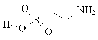
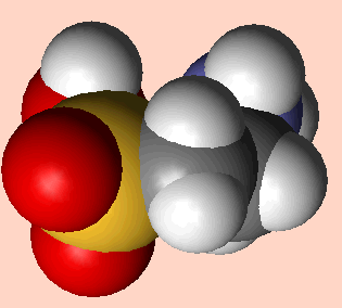
Taurine structure - click for 3D VRML structureTaurine - space fill model
Taurine - (2-aminoethane sulphonic acid) [1]

Its Role in Biology and Pharmacy

Taurine is synthesised by the human body, primarily in the liver by oxidation followed by decarboxylation of the amino acid cysteine.
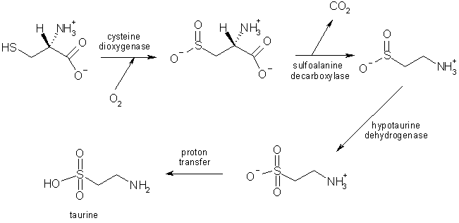
Biosynthesis of taurine
The biosynthesis of taurine from cysteine in the liver [5].

Taurine is one of a group of organic compounds which has been formed in experiments designed to simulate early-Earth conditions along with electrical discharges to simulate lightning. This result is often cited as evidence for the ease with which sulfur-containing biological molecules could be naturally synthesised under prebiotic conditions [6]. The conclusion is somewhat controversial, however, as the reaction occurred because of the presence of large concentrations of methane and ammonium hydroxide, the abundance of which on prebiotic Earth is questionable [7].

With a few exceptions, liver-synthesised taurine is not incorporated into polypeptides but is found free, lending it a unique set of properties, including a separate intercell transport mechanism and an independence from protein synthesis and catabolism [8]. Taurine has been linked to a wide range of bodily functions and Stapleton et al (1998) list roles in "osmoregulation, antioxidation, detoxification and stimulation of glycolysis and glycogenesis". The synthesis pathway of taurine is especially active in the early stages of life, and taurine is found in breast milk, suggesting that it is particularly vital at this stage. Taurine also plays a role in muscle contraction, where it enhances the ability of the muscles to generate force by catalysing the uptake and release of calcium ions [9].

Experiments on rats have also yielded evidence of a very substantial role in detoxification in the liver - Waters et al [10] report that high doses of taurine administered before or soon after ingestion of an overdose of paracetemol protected the livers of rats from hepatotoxcity and serious liver damage from the drugs. Although the idea has yet to progress to any form of human trial, the prophylactic and therapeutic benefits of the substance in cases of paracetemol overdose are promising .

A blind cat - a result of taurine deficiency?In fact, several areas of medical research are interested in taurine's pharmaceutical potential. For example, phase two clinical trials are currently underway at McLean Hospital in Massachusetts using taurine as an anti-manic agent to stabilise the mood of patients with bipolar disorder [11]. Tsuboyama-Kasaoka et al [12] also postulate a link between taurine deficiency and obesity in humans, showing that in mice, raising taurine levels in the body led to a greater resting rate of energy usage and less build-up of adipose tissue. The paper therefore suggests that taurine supplements may help prevent obesity.

Taurine in animals

Taurine is necessary for normal skeletal muscle functioning in humans and mice [13]. Cats cannot synthesise taurine, and so need it added as supplements in their diets. Without it, they suffer conditions such as blindness, hair loss and tooth decay. Taurine is also essential in the early development of many types of perching birds. Parent birds with new offspring often seek out spiders (which are rich in taurines) with which to feed their young. so, in this case, taurine really does give them wings!

References

1. Kim W., "Debunking the Effects of Taurine in Red Bull Energy Drink", Nutrition Bytes, 9, (2003) 6.

2. Seidl R. et al, "A Taurine and Caffeine-containing drink stimulates cognitive performance and well-being", Amino Acids, 19, (2000) 635.

3. Sharp D., Dictionary of Chemistry (Abridged), 3rd Edition, Penguin Books, 2003.

4. Stapleton P. et. al, "Host defense - a role for the amino acid taurine?", J. Parenteral and Enteral Nutrition, 22, (1998) 42.

5. Moss G., "Taurine Biosynthesis", http://www.chem.qmul.ac.uk/iubmb/enzyme/reaction/misc/taurine.html, Queen Mary University of London, 1992.

6. Ferris J., "Chemical markers of Prebiotic Chemistry in Hydrothermal systems", in Origin of Life and Evolution of Biospheres V.22 Numbers 1-4, ed. N Holm, Springer, Netherlands, Jan. 1992, p.120.

7. Wigley T., Nature, 291, (1981) 213.

8. Chiarla C. et al, "The Relationship between Plasma Taurine and Other Amino Acid Levels in Human Sepsis", J. of Nutrition, 130, (2000) 2222.

9. K. Harrison, "Taurine", http://www.3dchem.com/molecules.asp?ID=22, (2002).

10. Waters E. et al, "Role of taurine in preventing acetaminophen-induced hepatic injury in the rat", Am. J. Physiol. Gastrointest. Liver Physiol., 280, (2001) 1274.

11. Murphy B. et al, "Taurine as an Anti-Manic Agent", (2005) http://www.clinicaltrials.gov/ct/show/NCT00217165

12. Tsuboyama-Kasaoka et al, "Taurine (2-Aminoethanesulfonic Acid) Deficiency Creates a Vicious Circle Promoting Obesity", Endocrinology, 147, (2006) 3276.

13. Wikipedia - taurine

lunes, 9 de agosto de 2010

In a Video Game, Tackling the Complexities of Protein Folding

In a match that pitted video game players against the best known computer program designed for the task, the gamers outperformed the software in figuring out how 10 proteins fold into their three-dimensional configurations.

Proteins are essentially biological nano-machines that carry out myriad functions in the body, and biologists have long sought to understand how the long chains of amino acids that make up each protein fold into their specific configurations.

In May 2008, researchers at the University of Washington made a protein-folding video game called Foldit freely available via the Internet. The game, which was competitive and offered the puzzle-solving qualities of a game like Rubik’s Cube, quickly attracted a dedicated following of thousands of players.

The success of the Foldit players, the researchers report in this week’s issue of Nature, shows that nonscientists can collaborate to develop new strategies and algorithms that are distinct from traditional software solutions to the challenge of protein folding.

The researchers took pains to credit the volunteers who competed at Foldit in the last two years, listing “Foldit players” at the end of the report’s author list and noting that more than 57,000 players “contributed extensively through their feedback and gameplay.”

Zoran Popovic, a computer scientist at the University of Washington who was a lead author of the paper, said, “If things go according to plan, not too long from now, such massive author lists should be commonplace.” Foldit begins with a series of tutorials in which the player controls proteinlike structures on a computer display. In the game, as structures are modified, a score is calculated based on how well the protein is folded. Players are given a set of controls that let them do things like “shake,” “wiggle” and “rebuild” to reshape the backbone and the amino acid side shapes of a specific protein into a more efficient structure.

A list of top scores for each puzzle is posted so that players can compare their results. Players may also collaborate in teams, tracking progress on a separate list of group scores.

The protein-folding problem can be solved by computers using statistical and related software algorithms, but it takes an immense amount of processing power.

“The problem is that these proteins are far, far more complex than a robotic arm, and can ‘fold’ in time frames measured in billionths of a second,” Duke Ferris, founder of the GameRevolution Web site, wrote recently. “It’s like trying to solve a million-sided Rubik’s Cube while it also spins at 10,000 r.p.m. And that’s for just one ‘fold.’ ”

In a comparison involving 10 separate protein-folding puzzles, video game players matched the results generated by software solutions in three of the puzzles, outperformed them in five cases and found significantly better solutions in two others, according to the scientists.

In addition to the acuity of human pattern-recognition skills, the researchers noted that players outperformed the best software tools in other ways as well, writing: “Humans use a much more varied range of exploration methods than computers. Different players use different move sequences, both according to the puzzle type and throughout the duration of a puzzle.”

The Foldit project was inspired by the volunteers who were contributing the downtime on their home computers to power a protein-folding program called Rosetta@home. The computer donors could see the progress of the program on their screens, and they began to note inefficiencies in the software’s folding approach. That led the scientists to look for ways to systematically harness the skills of the human volunteers.

Two New Paths to the Dream: Regeneration

By NICHOLAS WADE Published: August 5, 2010

Two research reports published Friday offer novel approaches to the age-old dream of regenerating the body from its own cells.

Animals like newts and zebra fish can regenerate limbs, fins, even part of the heart. If only people could do the same, amputees might grow new limbs and stricken hearts be coaxed to repair themselves.

But humans have very little regenerative capacity, probably because of an evolutionary trade-off: suppressing cell growth reduced the risk of cancer, enabling humans to live longer. A person can renew his liver to some extent, and regrow a fingertip while very young, but not much more.

In the first of the two new approaches, a research group at Stanford University led by Helen M. Blau, Jason H. Pomerantz and Kostandin V. Pajcini has taken a possible first step toward unlocking the human ability to regenerate. By inactivating two genes that work to suppress tumors, they got mouse muscle cells to revert to a younger state, start dividing and help repair tissue.

What is true of mice is often true of humans, and although scientists are a long way from being able to cause limbs to regenerate, the research is attracting attention. Jeremy Brockes, a leading expert on regeneration at University College London, said the report was “an excellent paper.” Though there is a lot still to learn about the process, “it is hard to imagine that it will not be informative for regenerative medicine in the future,” he said.

In recent years, most research in the field of regenerative medicine has focused on the hope that stem cells, immature cells that give rise to any specific type of cell needed in the body, can somehow be trained to behave as normal adult cells do. Nature’s method of regeneration is quite different in that it starts with the adult cells at the site of a wound and converts the cells to a stemlike state in which they can grow and divide.

The Stanford team has taken a step toward mimicking the natural process. “What I like is that it’s built on what’s happening in nature,” Dr. Blau said. “We mammals lost this regenerative capacity in order to have better tumor suppression, but if we reawaken it in a careful way we could make use of it in a clinical setting.”

Dr. Pomerantz, a clinician, hopes the technique can be applied to people, though many more animal experiments need to be done first. “We have shown we can recapitulate in mammalian cells behavior of lower vertebrate cells that is required for regeneration,” he said. “We would propose using it in amputations of a limb or part of a limb or in cardiac muscle.” After a heart attack, the muscle cells do not regenerate, so any method of making them do so would be a possible treatment.

Interfering with tumor suppressor genes is a dangerous game, but Dr. Pomerantz said the genes could be inhibited for just a short period by applying the right dose of drug. When the drug has dissipated, the antitumor function of the gene would be restored.

Finding the right combination of genes to suppress was a critical step in the new research. One of the two tumor suppressor genes is an ancient gene, known as Rb, which is naturally inactivated in newts and fish when they start regenerating tissue. Mammals possess both the Rb gene and a backup, called the Arf gene, which will close down a cancer-prone cell if Rb fails to do so.

The Stanford team found that newts did not have the Arf backup gene, which mammals must have acquired after their lineage diverged from that of amphibians. This suggests that the backup system “evolved at the expense of regeneration,” the Stanford researchers say in Friday’s issue of Cell Stem Cell.

The Stanford team shut off both Rb and Arf with a chemical called silencing-RNA and found the mouse muscle cells started dividing. When injected into a mouse’s leg, the cells fused into the existing muscle fibers, just as they are meant to.

The Stanford researchers have learned how to block two genes thought to inhibit the natural regenerative capacity of cells, but it is somewhat surprising that the regenerative mechanism should still exist at all if mammals have been unable to use it for 200 million years. “One school of thought is that regeneration is a default mechanism and doesn’t require its own program,” Dr. Pomerantz said.

Dr. Brockes believes that this is true in part. Regeneration “depends on a largely conserved cellular machinery,” he said, meaning that it is present in all animals. The machinery comes into play in wound healing and tissue maintenance. But specific instances of regeneration, like regrowing a whole limb, are invoked by genes specific to various species. He has found a protein specific to salamanders that coordinates regrowth of a salamander limb.

If the regeneration of a whole limb is a special ability that salamanders have evolved, then humans would not have any inherent ability to do the same. “I would beware of suggesting that this sort of manipulation is capable of unlocking ‘the newt within,’ ” Dr. Brockes said.

A second, quite different approach to regenerating a tissue is reported in Friday’s issue of Cell by Deepak Srivastava and colleagues at the University of California, San Francisco. Working also in the mouse, they have developed a way of reprogramming the ordinary tissue cells of the heart into heart muscle cells, the type that is irretrievably lost in a heart attack.

The Japanese scientist Shinya Yamanaka showed three years ago that skin cells could be converted to embryonic stem cells simply by adding four proteins known to regulate genes. Inspired by Dr. Yamanaka’s method, Dr. Srivastava and his colleagues selected 14 such proteins and eventually found that with only three of them they could convert heart fibroblast cells into heart muscle cells.

To make clinical use of the discovery, Dr. Srivastava said he would need first to duplicate the process with human cells, and then develop three drugs that could substitute for the three proteins used in the conversion process. The drugs could be loaded into a stent, a small tube used in coronary bypass operations. With the stent inserted into a heart artery, the drugs would convert some of the heart’s tissue cells into heart muscle cells.

Some researchers hope that with Dr. Yamanaka’s method of turning skin cells into embryonic stem cells, those stem cells can be converted into usable heart muscle cells. One problem with this approach is that any unconverted embryonic stem cells may form tumors. Dr. Srivastava’s method sidesteps this problem by avoiding the stem cell stage.

martes, 3 de agosto de 2010

2 August 2010
BIOCHEMIST EVOLUTION
microRNA protects red blood cells from free radicals

Paediatric researchers have discovered a new biological pathway in which small segments of RNA, called microRNA, help protect red blood cells from injury caused by free radicals.

The microRNA seems to have only a modest role when red blood cells experience normal conditions, but steps into action when the cells are threatened by oxidant stress.

Led by haematologist Mitchell Weiss of The Children’s Hospital of Philadelphia, the current study describes how a particular microRNA fine-tunes gene activity by acting on an unexpected signalling pathway.

The study appears in the August 1 issue of the journal Genes & Development, simultaneously with a similar study of microRNAs and red blood cells by a University of Texas team led by Eric Olson. The two studies reinforce each other, said Weiss.

MicroRNAs are single-stranded molecules of ribonucleic acid (RNA) averaging only 22 nucleotides long. Scientists estimate that 500 to 1000 microRNAs exist in the human genome. First characterized in the early 1990s, they received their current name in 2001. Over the past decade, scientists have increasingly recognized that microRNAs play a crucial role in regulating genes, most typically by attaching to a piece of messenger RNA and blocking it from being translated into a protein, but many details remain to be discovered.

“Although microRNAs affect the formation and function of most or all tissues, for most microRNAs, we don’t know their precise mechanisms of action,” said Weiss. “In this case we already knew this microRNA, called miR-451, regulates red blood cells in zebrafish and mice, and because it is highly conserved in evolution, we presume it operates in humans as well. But its functional roles were poorly understood.”

By investigating how microRNAs influence red blood cell development, Weiss and colleagues aimed to understand how such development goes wrong in haemolytic anaemia, in which red blood cells are destroyed in large numbers, or in disorders of abnormal blood cell production. The current study used knockout mice — bioengineered animals in which the miR-451 gene was removed and could not function.

They found that preventing the activity of miR-451 produced only modest effects — mild anaemia in the mice — but when the team subjected mice to oxidant stress by dosing them with a drug that produces free radicals, the mice had profound anaemia. The oxygen radicals attacked haemoglobin, the iron-carrying molecule in red blood cells.

“This is a common theme in microRNAs — frequently, they don’t play a central role during tissue formation or normal conditions, but they have a strong protective effect when an organism is stressed,” said Weiss. “Over evolutionary time, red blood cells have evolved ways to protect themselves; one of those ways is the action of microRNA.”

Weiss’s team found that miR-451, acting through intermediate steps on a signalling pathway, affects a key protein, FoxO3. As a transcription factor, FoxO3 regulates hundreds of genes; in this case, FoxO3 stimulates specific genes that protect red blood cells from oxidant stress. The knockout mice in this study, having lost miR-451’s function, showed impaired FoxO3 activity, and less ability to protect their red blood cells.

The regulatory pathway seen here, Weiss added, may have medical implications beyond blood cell development. “This finding does not have immediate clinical application for patients with blood diseases, but it sheds light on how microRNAs fine tune physiological functions in different contexts,” said Weiss. FoxO3 regulates anti-oxidant functions in heart cells and also acts as a tumour suppressor, so miR-451 may have an important role in heart protection and in fighting cancers. “Further studies may broaden our knowledge of how this microRNA may defend the body against disease,” he added.

viernes, 25 de junio de 2010

DIABETES AND PREGNANCY 2011


Diabetes & Pregnancy Meeting Salzburg, Austria March 24-26, 2011

By embryology


The 6th International Symposium on Diabetes & Pregnancy (DIP) Salzburg, Austria March 24-26, 2011

The field of diabetes and pregnancy has come of age. From the conception of the terminology ‘gestational diabetes’ and ‘diabetes in pregnancy’ to the creation of an entire subspecialty, this Symposium documents the ‘gestation’ of the field.
This Symposium not only documents the past 80+ years of progress in the field of diabetes and pregnancy, but also presents the most up-to-date tools, techniques and management protocols to ensure the optimal outcome of pregnancies complicated by diabetes. In addition, the areas that remain controversial, such as screening and diagnosis, will be discussed in detail to enable participants to come to an opinion while waiting for the evidence to validate many of the expert opinions presented.
The latest theories and literature on the immunology of Type 1 diabetes will also be included, giving us hope that the near future holds the answers to prevention of this disease. Until there is a cure for diabetes, we must continually take on the weight of astutely diagnosing diabetes and treating all pregnant women who are at risk of an untoward outcome of pregnancy.
The faculty is comprised of renowned professionals and practitioners, who will present based on evidence as well as clinical experience.
In addition, this Symposium provides an international approach to enhancing the quality of care for women with diabetes in pregnancy, and answers for the clinician to enable delivery of optimal care for all pregnancies complicated by diabetes.

miércoles, 9 de junio de 2010

2010 International Year of Biodiversity

The UN has designated 2010 the 'International Year of Biodiversity’. To mark and celebrate this, the Society of Biology is organising a photographic competition. Entrants may submit one image per category. Entrants are free to interpret how they capture photographically images that celebrate, explore, comment on or reveal aspects of biodiversity from around the world.

Categories
  • Land – biodiversity associated with land
  • Air – biodiversity associated with the air
  • Water – biodiversity associated with water
  • Close up – biodiversity unseen by the naked eye
Prizes
  • Best in Show – £1,000
  • 1st prizes in each category – £250 in cash and Park Cameras vouchers
  • Top Young Photographer winner – £250 worth of vouchers
  • There will be additional prizes for runners up and certificates for ‘highly commended’ entries
The competition is free, open to all and will run between 20 April - 20 August 2010. Please review the competition rules before entering, details of the rules and entry forms are located to the right. Please contact photocomp@societyofbiology.org with any questions

Annual Symposium - Recent advances in membrane biochemistry

5—7 January 2011

Robinson College, Cambridge, UK


Earlybird registration deadline: 3 December 2010.

Registration will be available shortly.


Abstract submission deadline: 26 October 2010.

Abstract submission will be available shortly.


There are Student Travel Grants available for this meeting.

Oral communication slots are available at this meeting. All attendees, particularly young researchers, are invited to submit a poster abstract for consideration as an oral communication.


The proceedings of the meeting will be published as a stand-alone issue of Biochemical Society Transactions (online and in print) and a stand-alone volume of the online Symposia Series.

Symposium background
This symposium will highlight some of the recent advances in molecular membrane biochemistry. Membrane biology underpins a vast array of life processes such as bioenergetics, signalling and transport. In the last few years advances in this field has led to the development of large scale expression and purification of mammalian transmembrane proteins. This has then been paramount to enabling efficient crystallization and elucidation of the 3-dimensional structures of transmembrane proteins to occur.

In addition, other biophysical techniques have also been employed to further understand membrane protein structure, conformations, and interactions with other membrane components.


This symposia will focus on a number of key areas of current research advances, which will form the basis of 5 sessions:

1) Membrane protein expression and 3-dimensional structural analysis.


2) Examples of expression / structure analysis of membrane proteins which are of current interest.


3) Computer modelling of membrane protein structure, membrane protein folding and dynamics.


4) Lipid-protein interactions.

5) Membrane proteins in disease.

miércoles, 26 de mayo de 2010

Big Bang in the protein universe

Researchers of the Centre for Genomic Regulation (CRG) demonstrate evidence in support of the common ancestry of life thanks to a new computational approach to study protein evolution.

The study, published in Nature, reveals that protein evolution has not reached its limit and it is still continuing. At the same time, it provides us new information on why this evolution is so slow and conservative showing that protein structures are more evolutionary plastic than previously thought.

Almost 100 years ago Edwin Hubble observed that distant galaxies are moving away from Earth faster than those that are closer. This relationship between distance and velocity is widely cited as evidence of the origin of the Universe from a Big Bang. Researchers at the Centre for Genomic Regulation copied his approach to investigate the divergence between protein sequences.

“We wanted to know if the divergent evolution between proteins was still proceeding. Today, we can find proteins that are still similar after almost 3.5 billion years of evolution. Our study showed that their divergence continues with these proteins becoming more and more different despite their incredible level of conservation”, said Fyodor Kondrashov, principal investigator of the project and leader of the Evolutionary Genomics group at the CRG.

The work done by Fyodor Kondrashov and Inna Povolotskaya goes beyond similarity studies and discusses the evolution of proteins from the view of evolutionary dynamics, offering a new perspective on how protein structures are maintained in evolution. “In the same way that Hubble’s observations led to an understanding of the past and the future of our universe, using his approach at a molecular level we get a similar overview that gives us the ability to analyze evolutionary dynamics and get a broad prediction of the possible changes to the proteins in the future”, says Inna Povolotskaya, first author of the work and responsible for obtaining and analyzing all data.

Proteins are formed through combinations of amino acids, with only 20 types of amino acids are available to form a particular protein. To obtain the data for their study, the CRG researchers have compared proteins sequences from different species that were available in GenBank, a public database of genetic information. Comparing these sequences the authors measured the distance of proteins from each other and devised a method for measuring how fast the proteins are accumulating different changes. Thus, they could replicate Hubble’s approach by correlating the distance between the proteins with the rate of their divergence. The result indicates that even the most distantly-related proteins are still accumulating differences.

The study shows how new techniques of bioinformatics and computational analysis can also expand knowledge at a molecular level. “Our work is a good example of how we can learn new and very fundamental things just by analyzing a larger volume of data that can be obtained by one experimental laboratory”, says Kondrashov.

Most changes in a protein are deleterious because they somehow disrupt its structure or function. The authors observation that even very conservative proteins are still diverging challenges this view, because it implies that most amino acids in a protein can be changes without any ill effects. Their explanation is that amino acid changes that are deleterious in one combination can be benign when occurring in a different one. “Thanks to our study we now have a better understanding of protein structure dynamics” said Kondrashov.

The work of Povolotskaya and Kondrashov also provides new information on how different interactions between different amino acids in the structure of proteins slows down but does not completely prevent evolution.

Tomado de: http://www.biochemist.org/news/page.htm?item=40125